Question: High-resolution 2-D electrophoresis
(DE) proteomic analysis of atrial tissue. Representative 2-DE…
High-resolution 2-D electrophoresis
(DE) proteomic analysis of atrial tissue. Representative 2-DE gel
images corresponding to atrial
tissue proteins from patients with atrial fibrillation (A) and
sinus rhythm (B). The pI range is 4–7. The figure shows the
location on the 2D gels of the differentially regulated protein
features.
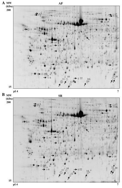
The analysis allowed the detection of
over 2300 protein spots per gel. Following differential image
analysis, 22 protein spot differences were found between the AF and
SR groups in the 4–7 isoelectric point range, leading to the
identification of 15 differentially regulated proteins. The main
group of proteins identified was that of heat shock proteins. Give
protein numbers for those down-regulated in atrial tissue from AF
patients?