Transcribed Image Text from this Question
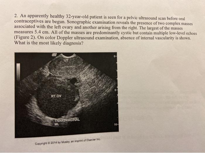
2. An apparently healthy 32-year-old patient is seen for a pelvic ultrasound scan before oral contraceptives are begun. Sonographic examination reveals the presence of two complex masses associated with the left ovary and another arising from the right. The largest of the masses measures 5.4 cm. All of the masses are predominantly cystic but contain multiple low-level echoes (Figure 2). On color Doppler ultrasound examination, absence of internal vascularity is shown. What is the most likely diagnosis? 0- UT RT OV TRANSVAGINAL ML 10 Copyright 2014 by Mosby, an imprint of Elsevier Inc.
(Visited 5 times, 1 visits today)