Transcribed Image Text from this Question
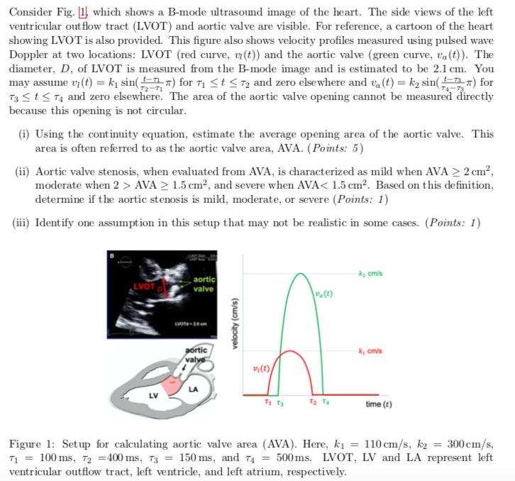
Consider Fig. 1), which shows a B-mode ultrasound image of the heart. The side views of the left ventricular out flow tract (LVOT) and aortic valve are visible. For reference, a cartoon of the heart showing LVOT is also provided. This figure also shows velocity profiles measured using pulsed wave Doppler at two locations: LVOT (red curve, vr(t)) and the aortic valve (green curve, va(t)). The diameter, D. of LVOT is measured from the B-mode image and is estimated to be 2.1 cm. You may assume vi(t) = k sin() for 71 St <72 and zero elsewhere and valt) = k2 sin() for 13<t<T4 and zero elsewhere. The area of the aortic valve opening cannot be measured directly because this opening is not circular. (i) Using the continuity equation, estimate the average opening area of the aortic valve. This area is often referred to as the aortic valve area, AVA. (Points: 5) (ii) Aortic valve stenosis, when evaluated from AVA, is characterized as mild when AVA > 2 cm², moderate when 2 > AVA > 1.5 cm², and severe when AVA< 1.5 cm². Based on this definition, determine if the aortic stenosis is mild, moderate, or severe (Points: 1) (iii) Identify one assumption in this setup that may not be realistic in some cases. (Points: 1) cmis LVOT DU aortic valve (0) LOT-28cm velocity (cm/s) aortic valvo cm/s 17.() Тата time (e) Figure 1: Set up for calculating aortic valve area (AVA). Here, ki = 110 cm/s, k2 = 300cm/s, T1 = 100 ms, T2 =400 ms, T3 = 150 ms, and T4 = 500 ms. LVOT, LV and LA represent left ventricular outflow tract, left ventricle, and left atrium, respectively.
(Visited 4 times, 1 visits today)

 help with i and ii please!!

