Transcribed Image Text from this Question
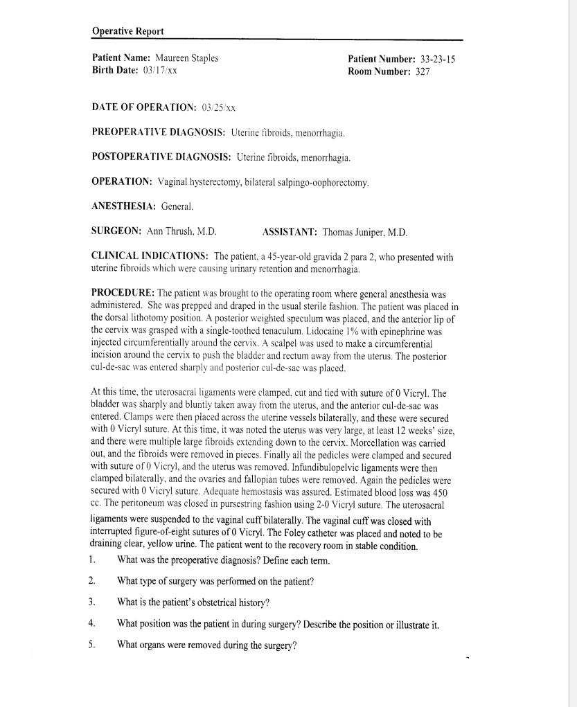
Operative Report Patient Name: Maureen Staples Birth Date: 03/17/XX Patient Number: 33-23-15 Room Number: 327 DATE OF OPERATION: 03/25/XX PREOPERATIVE DIAGNOSIS: Uterine fibroids, menorrhagia. POSTOPERATIVE DIAGNOSIS: Uterine fibroids, menorrhagia. OPERATION: Vaginal hysterectomy, bilateral salpingo-oophorectomy. ANESTHESIA: General. SURGEON: Ann Thrush. M.D. ASSISTANT: Thomas Juniper, M.D. CLINICAL INDICATIONS: The patient, a 45-year-old gravida 2 para 2, who presented with uterine fibroids which were causing urinary retention and menorrhagia. PROCEDURE: The patient was brought to the operating room where general anesthesia was administered. She was prepped and draped in the usual sterile fashion. The patient was placed in the dorsal lithotomy position. A posterior weighted speculum was placed, and the anterior lip of the cervix was grasped with a single-toothed tenaculum. Lidocaine 1% with epinephrine was injected circumferentially around the cervix. A scalpel was used to make a circumferential incision around the cervix to push the bladder and rectum away from the uterus. The posterior cul-de-sac was entered sharply and posterior cul-de-sac was placed. At this time, the uterosacral ligaments were clamped, cut and tied with suture of O Vicryl. The bladder was sharply and bluntly taken away from the uterus, and the anterior cul-de-sac was entered. Clamps were then placed across the uterine vessels bilaterally, and these were secured with 0 Vicryl suture. At this time, it was noted the uterus was very large, at least 12 weeks’ size, and there were multiple large fibroids extending down to the cervix. Morcellation was carried out, and the fibroids were removed in pieces. Finally all the pedicles were clamped and secured with suture of O Vicryl, and the uterus was removed. Infundibulopelvic ligaments were then clamped bilaterally, and the ovaries and fallopian tubes were removed. Again the pedicles were secured with 0 Vicryl suture. Adequate hemostasis was assured. Estimated blood loss was 450 cc. The peritoneum was closed in pursestring fashion using 2-0 Vicryl suture. The uterosacral ligaments were suspended to the vaginal cuff bilaterally. The vaginal cuff was closed with interrupted figure-of-eight sutures of Vicryl. The Foley catheter was placed and noted to be draining clear, yellow urine. The patient went to the recovery room in stable condition. 1. What was the preoperative diagnosis? Define each term. 2. What type of surgery was performed on the patient? 3. What is the patient’s obstetrical history? 4. What position was the patient in during surgery? Describe the position or illustrate it. 5. What organs were removed during the surgery?
(Visited 3 times, 1 visits today)


