Transcribed Image Text from this Question
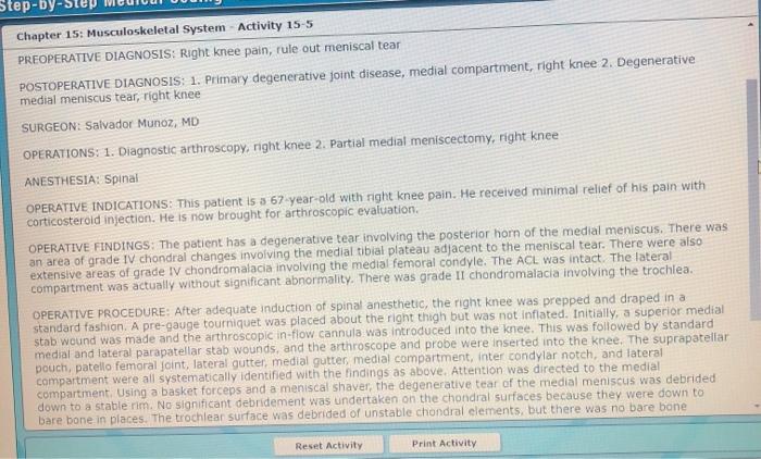
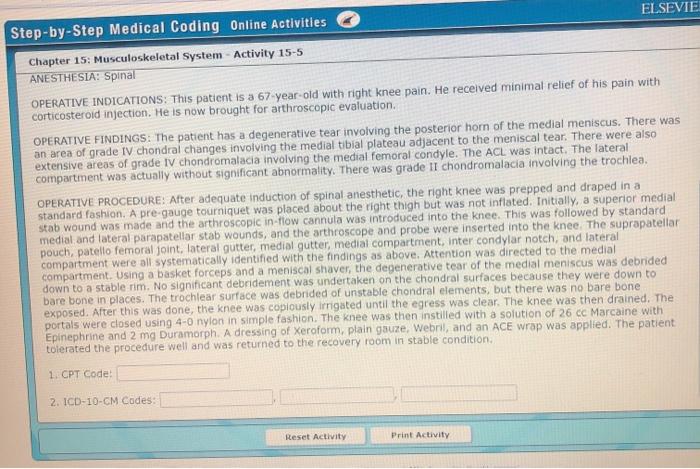
Step-by-ste Chapter 15: Musculoskeletal System Activity 15-5 PREOPERATIVE DIAGNOSIS: Right knee pain, rule out meniscal tear POSTOPERATIVE DIAGNOSIS: 1. Primary degenerative joint disease, medial compartment, right knee 2. Degenerative medial meniscus tear, right knee SURGEON: Salvador Munoz, MD OPERATIONS: 1. Diagnostic arthroscopy, right knee 2. Partial medial meniscectomy, right knee ANESTHESIA: Spinal OPERATIVE INDICATIONS: This patient is 3 67-year-old with right knee pain. He received minimal relief of his pain with corticosteroid injection. He is now brought for arthroscopic evaluation OPERATIVE FINDINGS: The patient has a degenerative tear involving the posterior horn of the medial meniscus. There was an area of grade IV chondral changes involving the medial tibial plateau adjacent to the meniscal tear. There were also extensive areas of grade ly chondromalacia involving the medial femoral condyle. The ACL was intact. The lateral compartment was actually without significant abnormality. There was grade II chondromalacia involving the trochlea. OPERATIVE PROCEDURE: After adequate induction of spinal anesthetic, the right knee was prepped and draped in a standard fashion. A pre-gauge tourniquet was placed about the right thigh but was not inflated. Initially, a superior medial stab wound was made and the arthroscopic in-flow cannula was introduced into the knee. This was followed by standard medial and lateral parapatellar stab wounds, and the arthroscope and probe were inserted into the knee. The suprapatellar pouch, patello femoral joint, lateral gutter, medial gutter, medial compartment, inter condylar notch, and lateral compartment were all systematically identified with the findings as above. Attention was directed to the medial compartment. Using a basket forceps and a meniscal shaver, the degenerative tear of the medial meniscus was debrided down to a stable nim. No significant debridement was undertaken on the chondral surfaces because they were down to bare bone in places. The trochlear surface was debrided of unstable chondral elements, but there was no bare bone Reset Activity Print Activity Step-by-Step Medical Coding Online Activities ELSEVIE Chapter 15: Musculoskeletal System – Activity 15-5 ANESTHESIA: Spinal OPERATIVE INDICATIONS: This patient is a 67-year-old with right knee pain. He received minimal relief of his pain with corticosteroid injection. He is now brought for arthroscopic evaluation OPERATIVE FINDINGS: The patient has a degenerative tear involving the posterior hom of the medial meniscus. There was an area of grade IV chondral changes involving the medial tibial plateau adjacent to the meniscal tear. There were also extensive areas of grade IV chondromalacia involving the medial femoral condyle. The ACL was intact. The lateral compartment was actually without significant abnormality. There was grade II chondromalacia involving the trochlea. OPERATIVE PROCEDURE: After adequate induction of spinal anesthetic, the right knee was prepped and draped in a standard fashion. A pre-gauge tourniquet was placed about the right thigh but was not inflated. Initially, a superior medial stab wound was made and the arthroscopic in-flow cannula was introduced into the knee. This was followed by standard medial and lateral parapatellar stab wounds, and the arthroscope and probe were inserted into the knee The suprapatellar pouch, patello femoral joint, lateral gutter, medial gutter, medial compartment, inter condylar notch, and lateral compartment were all systematically identified with the findings as above. Attention was directed to the medial compartment. Using a basket forceps and a meniscal shaver, the degenerative tear of the medial meniscus was debrided down to a stable rim. No significant debridement was undertaken on the chondral surfaces because they were down to bare bone in places. The trochlear surface was debrided of unstable chondral elements, but there was no bare bone exposed. After this was done, the knee was copiously irrigated until the egress was clear. The knee was then drained. The portals were closed using 4-0 nylon in simple fashion. The knee was then instilled with a solution of 26 cc Marcaine with Epinephrine and 2 mg Duramorph. A dressing of Xeroform, plain gauze, webril, and an ACE wrap was applied. The patient tolerated the procedure well and was returned to the recovery room in stable condition 1. CPT Code: 2. ICD-10-CM Codes: Reset Activity Print Activity
(Visited 11 times, 1 visits today)