Question: The plasmid diagrammed above contains three sites for the restriction enzyme. PstI, as shown. The…
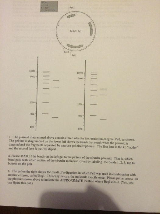
Show transcribed image text The plasmid diagrammed above contains three sites for the restriction enzyme. PstI, as shown. The gel that is diagrammed on the lower left shows the bands that result when the plasmid is digested and the fragments separated by agarose gel electrophorsis. The first lane is the kb "ladder" and the second lane is the PstI digest. Please MATCH the bands on the left gel.o the picture of the circular plasmid That is which band goes with which section of the circular molecule. (start by labeling the bands 1, 2, 3, top to bottom on the gel). The gel on the right shows the result of a digestion in which PstI was used in combination with another enzyme, called Bcgl. This enzyme cuts the molecule exactly once plrase put an arrow on the plasmid shown above to indicate the APPROXIMATE location where Bcgl cuts it. (yes,you can figure this out.)
The plasmid diagrammed above contains three sites for the restriction enzyme. PstI, as shown. The gel that is diagrammed on the lower left shows the bands that result when the plasmid is digested and the fragments separated by agarose gel electrophorsis. The first lane is the kb "ladder" and the second lane is the PstI digest. Please MATCH the bands on the left gel.o the picture of the circular plasmid That is which band goes with which section of the circular molecule. (start by labeling the bands 1, 2, 3, top to bottom on the gel). The gel on the right shows the result of a digestion in which PstI was used in combination with another enzyme, called Bcgl. This enzyme cuts the molecule exactly once plrase put an arrow on the plasmid shown above to indicate the APPROXIMATE location where Bcgl cuts it. (yes,you can figure this out.)